
Fig. 8
- ID
- ZDB-FIG-150319-12
- Publication
- Takeuchi et al., 2015 - Establishment of Gal4 transgenic zebrafish lines for analysis of development of cerebellar neural circuitry
- Other Figures
- All Figure Page
- Back to All Figure Page
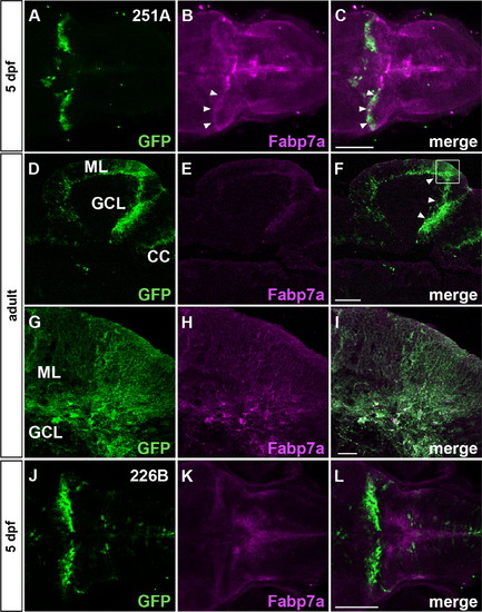
Bergmann glia-specific GFF lines. The SAGFF(LF)251A and SAGFF(LF)226B lines were crossed with UAS:GFP. (A–I) SAGFF(LF)251A; UAS:GFP. (J–L) SAGFF(LF)226B; UAS:GFP. The 5-dpf larvae (A–C, J–L, dorsal projection views with anterior to the left) and sagittal sections of adult brains (D–F, anterior to the left) were stained with anti-GFP (green, A, C, D, F, G, I, J, L) and anti-Fabp7 (magenta, B, C, E, F, H, I, K, L) antibodies. (G–I) High magnification views of the box in F. Note that GFP and Fabp7a staining colocalized only in the cerebella of larvae and adults (marked by arrowheads, B, C, F), indicating that SAGFF(LF)251A; UAS:GFP was expressed only in Bergmann glial cells, and not in the radial glia of other brain regions. SAGFF(LF)226B; UAS:GFP-positive signals were detected in the Bergmann glia of larvae, but not adult cerebella. Scale bars: 100 µm in C (applied to A-C); 200 µm in F (applied to D–F); 20 µm in I (applied to G–I); 100 µm in L (applied to J-L). |
| Gene: | |
|---|---|
| Antibody: | |
| Fish: | |
| Anatomical Term: | |
| Stage Range: | Day 5 to Adult |
Reprinted from Developmental Biology, 397(1), Takeuchi, M., Matsuda, K., Yamaguchi, S., Asakawa, K., Miyasaka, N., Lal, P., Yoshihara, Y., Koga, A., Kawakami, K., Shimizu, T., Hibi, M., Establishment of Gal4 transgenic zebrafish lines for analysis of development of cerebellar neural circuitry, 1-17, Copyright (2015) with permission from Elsevier. Full text @ Dev. Biol.