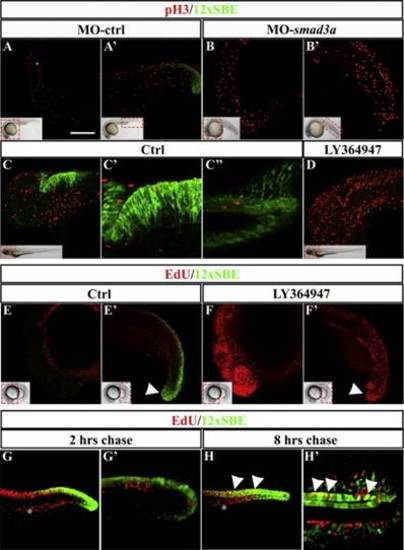
Smad3/TGFβ signaling is mainly active in post-mitotic cells. (A–B′) Confocal lateral views of immunofluorescence for GFP (green) and phospho-Histone3 (pH3, red) on Tg(12xSBE:EGFP)ia16 embryos injected at 1–2 cell stage either with the control (A), or smad3a-morpholino (B). For each confocal picture (Z-stack), a small brightfield view of a morphant embryo shows which area is displayed (red dashed line). (C–D) Confocal lateral views of immunofluorescence for GFP (green) and pH3 (red) on 2 dpf larva of Tg(12xSBE:EGFP)ia16 line treated with either DMSO (C) or LY364947 (D) at 24hpf. For each confocal picture (Z-stack), a small brightfield view of a 48 hpf larva shows which area is displayed (red dashed line). (C′ and C′′) Confocal zoomed views (single plane) of hindbrain and tail, respectively, of immunofluorescence for GFP (green) and pH3 (red) on DMSO-treated larva of the 12xSBE line. (E–F) Confocal lateral views of the head (E, F) or tail (E′, F′) regions after EdU labeling on 20 hpf embryos treated at 12 hpf either with DMSO (E, E′) or LY364947 (F, F′). For each confocal picture (Z-stack), a small brightfield view of a morphant embryo shows which area is displayed (red dashed line). (G, H) confocal lateral images (Z-stack) of pulse and chase EdU assay on 24 hpf embryos of Tg(12xSBE:EGFP)ia16 line. Embryos have been fixed after a chase of either 2 (G–G′) or 8 (H–H′) hours (hrs) and immunostained for EdU (red) and GFP (green). EdU+ cells fixed after 2 h are roughly in S/G2 phase, while EdU+ cells fixed after 8 h of chase are post-mitotic. (G′, H′) Zoomed view (single plane) of Tg(12xSBE:EGFP)ia16 tail after 2 or 8 h chase. Scale bar is 100 µm in A–B′, E–F′, G, H; 50 µm in C′–C′&prime& 20 µm in G′, H′. Quantification is presented in Supplemental Table S4.
|
