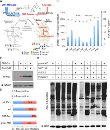
slc35c1 enhances the level of N-linked fucosylation expression in zebrafish embryos. (A) Schematic illustration of fucose biosynthetic pathways. Slc35c1 transports GDP-Fuc into the late secretary pathway (mainly the Golgi apparatus). (B) slc35c1 mRNA expression during zebrafish development as quantified by qPCR. The blue bars show slc35c1 relative expression during development and the red dots show the qPCR cycle number of the control gene odc1. (C) Western blot analysis of lysates of 5hpf zebrafish embryos expressing mouse Slc35c1-myc fusion protein. (D) Western blot analysis of global fucosylation levels at shield stage (6hpf) in embryos microinjected with gmds MO or mouse slc35c1-myc mRNA in the presence or absence of GDP-Fuc before and after PNGase F digestion. Proteins were resolved by SDS-PAGE and IB with AAL followed by anti-biotin-HRP in (D) or by streptavidin-Alexa488 for the quantification in (E). N-fucosylation: N-linked fucosylation; O-fucosylation: fucose in mucin and O-fucosylated proteins.
|
