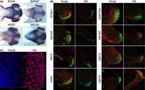
In situ hybridization and immunohistochemistry. (A) Dorsal view of 72 hpf zebrafish embryos processed by in situ hybridization for ZNF367, KRCP, and MEX3A. Proliferating cell nuclear antigen (PCNA) riboprobe has been used to visualize proliferative region of the zebrafish larval brain in the optic tectum (green arrowhead) and retina (red arrow head). (B) Double-labeling in situ hybridization (ISH) and immunohistochemistry (IHC) on the posterior margin of the optic tectum (OTp) in Nothobranchius furzeri for the following genes: SCML4, MEX3A, KRCP, AGR2, DNMT3AA, ZNF367, CBX1A, and CBX7A – ISH signal was revealed using fluorescent Fast Red and is visualized in red, PCNA IHC is visualized in green to localize the proliferative niche of the OTp. Only merged signals are shown. Young animals were 6 weeks old, and old animals were 25 weeks old. Separated red and green channels are reported in Fig. S10. The images are representative of three replicates. (C) Changes of PRC2 markers with age. Immunohistochemical detection of H3K27me3 in the posterior optic tectum of young (5 weeks) and old (35 weeks) individuals. The images are representative of three replicates. Immunohistochemical signal is shown in red and DNA staining (TOPRO3) in blue. Overlap of signals gives rise to a purple hue.
|
