
Fig. 1
- ID
- ZDB-FIG-150213-1
- Publication
- Paik et al., 2013 - A cdx4-sall4 regulatory module controls the transition from mesoderm formation to embryonic hematopoiesis
- Other Figures
- All Figure Page
- Back to All Figure Page
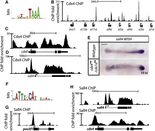
Cdx4 and Sall4 Bind to Each Other’s Genomic Locus (A) Cdx4-binding sites are enriched for the ATAAA motif. The most enriched motif was identified using MEME (Bailey and Elkan, 1994). (B) Gene track of the hox aa locus, showing Cdx4 binding at genomic regions along the x axis and the total number of reads on the y axis. (C) Gene track of the cdx4 locus, showing Cdx4 binding to its own locus. (D) Gene track of the sall4 locus, showing Cdx4 binding to sall4 locus. (E) WISH of sall4 mRNA expression level in 10-somite stage wild-type embryos and cdx4mo/cdx1amo. Scale bar = 200 µm. (F) Sall4-binding sites are enriched for the ATTTGCAT motif, an Oct4 motif. (G) Gene track of the pou5f1 locus, showing Sall4 binding. (H) Gene track of sall4 and cdx4 loci, showing Sall4 binding. See also Figure S1. |
| Gene: | |
|---|---|
| Fish: | |
| Knockdown Reagents: | |
| Anatomical Term: | |
| Stage: | 10-13 somites |