
Fig. S5
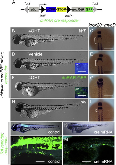
Over-expression of dominant-negative zebrafish RA receptor α inhibits endogenous RA signaling. (A) Schematic of the dnRAR cre responder, Tg(ubb:loxP-eCFP-loxP-dnRAR-GFP)jh39. Tol2 arms (gray triangles) flank the whole expression cassette. Ubb promoter/enhancer drives ubiquitous expression. Active cre induces recombination between the two loxP sites (black triangles), removing the STOP signal (yellow box), and allowing production of the dnRAR-GFP fusion protein. (B, D, F, and H) Shows phase contrast images of whole 3 dpf larvae. (D′ and F′) Fluorescent images of 3 dpf larval trunk. Before recombination cells are labeled with eCFP (blue) and after cre activity with nuclear GFP (green). Scale Bar, 25 μm. (C, E, G, and I) Dorsal views of 11-somite stage embryos following whole mount in situ hybridization to detect krox20 and myoD expression. Scale Bar 200 μm. Different experimental conditions as follows: (B and C) wild-type fish incubated from 4–72 hpf with 4OHT. (D–G) Fish transgenic for the ubiquitous creERT2driver and the dnRAR cre responder, were treated with vehicle (D and E) or 4OHT (F and G) from 4–72 hpf. For phenotypic comparison to larvae lacking ALDH1a2 activity, ALDH1a2nls/nls mutant larvae were analyzed (H and I). After 4OHT treatment, unlike controls (B and D), larvae transgenic for the ubiquitous creERT2driver and dnRAR cre responder have nuclear localized GFP, due to the expression of dnRAR-GFP (F′), and a gross morphological phenotype (F) similar to the nls mutant (H). The deleterious phenotype associated with compromised RA signaling includes patterning defects (G and I) when compared to controls (C and E). These defects can be visualized by a decrease in distance (white bracket) between the most anterior somite (marked by myoD expression) and rhombomere 5 (marked by krox20 expression) (Oxtoby and Jowett, 1993; Weinberg et al., 1996). (J–M) Fluorescent images of the larvae of dnRAR cre responder; RA reporter at 72 hpf. Cre mRNA was injected at one-cell stage to induce widespread expression of dnRAR-GFP fusion protein (K and M), while non-injected larvae were used as controls (J and L). Cytoplasmic-GFP signal (clearly seen in L) indicates the activity of RA reporter, and nuclear-GFP signal (visible in M) indicates the induction of dnRAR-GFP fusion protein. Induction of dnRAR-GFP greatly diminishes activity of the RA-reporter. Scale bar, 50 μm in L and M. |
Reprinted from Developmental Biology, 394(1), Huang, W., Wang, G., Delaspre, F., Vitery, M.D., Beer, R.L., Parsons, M.J., Retinoic acid plays an evolutionarily conserved and biphasic role in pancreas development, 83-93, Copyright (2014) with permission from Elsevier. Full text @ Dev. Biol.