
Fig. 4
- ID
- ZDB-FIG-141125-5
- Publication
- Choksi et al., 2014 - Systematic discovery of novel ciliary genes through functional genomics in the zebrafish
- Other Figures
- All Figure Page
- Back to All Figure Page
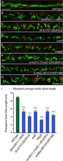
FIGs are required for both ciliogenesis and cilia organization. (A-H) Cilia of the pronephric duct were stained with anti-Arl13b antibodies (green) and the basal bodies were stained with antibodies to γ-tubulin (red) in 24hpf embryos. (A) Long cilia of uniform length and orientation are visible in a wild-type embryo. (B-G) Six morphants exhibited shortening of pronephric cilia at 24hpf. (H) The knockdown of mustn1a caused curling of cilia (arrows) and a disorganization of the γ-tubulin (arrowheads). Scale bar: 10μm. (I) Measurements of ciliary length in wild-type versus morphant embryos for six genes reveals the extent of ciliary shortening in the morphants. Error bars represent s.e.m. ***P<5.0×10⁷ (Student′s t-test, two-tailed, P-values are listed in supplementary material Table S3); n≥30. |
| Fish: | |
|---|---|
| Knockdown Reagents: | |
| Observed In: | |
| Stage: | Prim-5 |