
Fig. 3
- ID
- ZDB-FIG-140807-11
- Publication
- Kurtenbach et al., 2013 - Pannexin1 channel proteins in the zebrafish retina have shared and unique properties
- Other Figures
- All Figure Page
- Back to All Figure Page
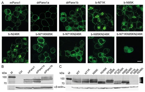
(A) Localization of Panx1-EYFP WT and glycosylation-deficient drPanx1b-EYFP mutant proteins in N2a cells. Protein-derived fluorescence was detected using confocal laser scanning microscopy 48 h post transfection. (Scale bar = 10 µm) (B) Western blot analyses of Panx1-EYFP WT and (C) glycosylation-deficient drPanx1b-EYFP mutants. N2a cells transiently transfected with pEYFP-N1 expression vector constructs were lysed 48 h post transfection. 20 µg total protein were subjected to SDS-PAGE and subsequent western blot analyses using the mouse anti-GFP and anti-mouse IRDye680RD antibodies (B) or rabbit anti-GFP and anti-rabbit IRDye680LT for detection of the Panx1 fusion proteins (C). Mouse anti-β-actin served as a control for equal protein loading and was detected using anti-mouse IRDye680RD (B) or anti-mouse IRDye800CW (C). Arrowheads in (C) indicate the six drPanx1b bands. |