
FIGURE
Fig. 1
- ID
- ZDB-FIG-140804-35
- Publication
- Sanders et al., 2013 - Verification of Intraovum Transmission of a Microsporidium of Vertebrates: Pseudoloma neurophilia Infecting the Zebrafish, Danio rerio
- Other Figures
- All Figure Page
- Back to All Figure Page
Fig. 1
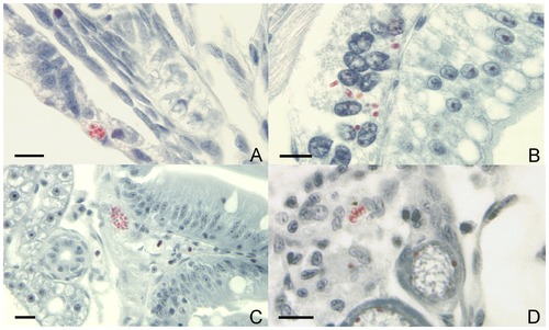
Spores of Pseudoloma neurophilia in Luna-stained histological sections of progeny of infected zebrafish, Danio rerio. A.Spores (red) in the epidermis of a 7 d post-fertilization (pf) larval zebrafish. B. Spores in the resorbing yolk-sac of the same 7 dpf larval zebrafish. C. Spore aggregate beneath the intestinal epithelium of an 8 wk pf juvenile fish. D. Spores in the ovigerous stroma adjacent to developing follicles in an 8 wk pf fish. Bar = 10 μm. |
Expression Data
Expression Detail
Antibody Labeling
Phenotype Data
Phenotype Detail
Acknowledgments
This image is the copyrighted work of the attributed author or publisher, and
ZFIN has permission only to display this image to its users.
Additional permissions should be obtained from the applicable author or publisher of the image.
Full text @ PLoS One