
Fig. S1
- ID
- ZDB-FIG-140728-19
- Publication
- Acosta et al., 2014 - Mutant Human FUS Is Ubiquitously Mislocalized and Generates Persistent Stress Granules in Primary Cultured Transgenic Zebrafish Cells
- Other Figures
- All Figure Page
- Back to All Figure Page
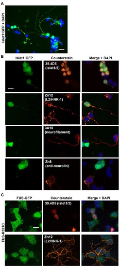
Zebrafish cell culturing protocol supports the growth and differentiation of motor neurons. (A) Cell cultures from transgenic zebrafish embryos expressing GFP under the motor neuron promoter islet-1 (islet1: GFP) demonstrated that motor neurons represented <10% of the cells in culture and exhibited extensive differentiation with axonal growth and branching (arrow). (B) Zebrafish neural cell-associated antibodies obtained from the Developmental Studies Hybridoma Bank (University of Iowa) were used: 39.4D5 [anti-islet-1/2] – primary motor neuron-specific transcription factor; Zn12 [anti-L2/HNK-1] – neural cell adhesion molecule (labels many different neural subtypes); 3A10 [anti-neurofilament] - derived from a neurofilament-associated antigen and labels a subset of hindbrain spinal cord projecting neurons such as Mauthner neurons (Brand et al. 1996) but appears not to label islet 1/2 expressing motor neurons; Zn8 [anti-neurolin] - expressed by secondary but not primary motoneurons during zebrafish development. This labeling demonstrated a mix of different neural subtypes in the cultures. (C) FUS-GFP was expressed in the cell soma of motor neurons and was not extensively transported into neurites. Scale bar = 20 μm. |