
Fig. 9
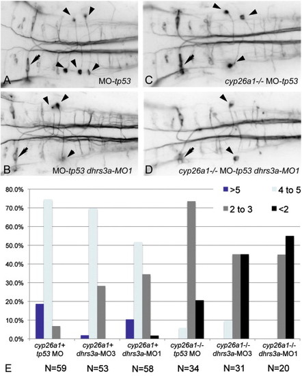
Dhrs3 knockdown effects hindbrain neuronal organization. (A–D) RMO44 immunostaining of hindbrain reticulospinal neurons: (A) control wt embryo with 5 ng tp53 MO; (B) dhrs3a MO1+tp53 MO-injected embryo; (C) cyp26a1 mutant embryo injected with 5 ng control tp53 MO; (D) cyp26a1 mutant embryo injected with dhrs3a MO1+tp53 MO. Arrows indicate the Mauthner neuron in r4, which is unaffected under all conditions; arrowheads indicate the distinctive T-interneurons which lie in r7 and anterior r8. (E) Quantification of the number of T-interneurons in each condition. N refers to the number of embryos counted. |
| Antibody: | |
|---|---|
| Fish: | |
| Knockdown Reagents: | |
| Anatomical Terms: | |
| Stage: | 1-4 somites |
| Fish: | |
|---|---|
| Knockdown Reagents: | |
| Observed In: | |
| Stage: | 1-4 somites |
Reprinted from Developmental Biology, 338(1), Feng, L., Hernandez, R.E., Waxman, J.S., Yelon, D., and Moens, C.B., Dhrs3a regulates retinoic acid biosynthesis through a feedback inhibition mechanism, 1-14, Copyright (2010) with permission from Elsevier. Full text @ Dev. Biol.