
Fig. 7
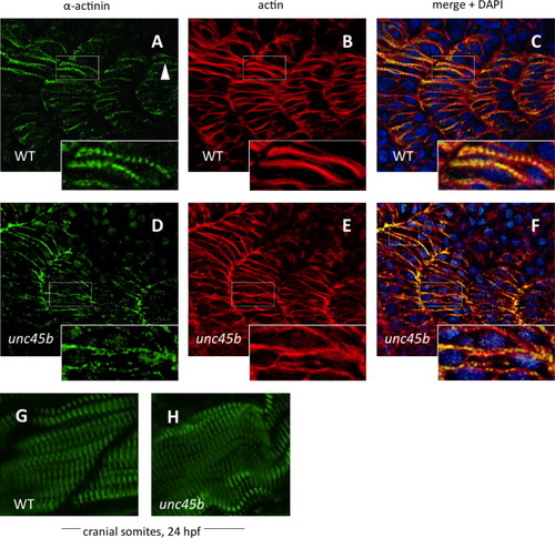
Nucleation of α-actinin is delayed in zebrafish unc45b mutants. Immunofluorescent staining of WT (A–C) and unc45b mutant embryos (D–F) at 20 h post-fertilization, displaying the 19th to 22nd somites. The caudal-most somites of these embryos were still undergoing myogenesis, as shown by the incomplete elongation of myofibers and localization of α-actinin and actin staining in WT embryos (A and B, inserts). Nucleation of α-actinin is the first indication of periodic myofibril patterning (arrowhead). In mutant embryos, organization of α-actinin at costamere attachment sites was not yet complete, and nucleation had just begun (D, insert). Actin counter-staining with phalloidin (B, E) demonstrates the ongoing organization of actin in early myofibers, which can be compared with the pattern of α-actinin localization in merged images (C, F). Blue fluorescence indicates DAPI nuclear stain. The cranial-most somites at 24 hpf display relatively normal patterns of α-actinin staining in mutants (H) compared to WT embryos (G). |
| Antibody: | |
|---|---|
| Fish: | |
| Anatomical Term: | |
| Stage: | 20-25 somites |
| Fish: | |
|---|---|
| Observed In: | |
| Stage: | 20-25 somites |
Reprinted from Developmental Biology, 390, Myhre, J.L., Hills, J.A., Jean, F., Pilgrim, D.B., Unc45b is essential for early myofibrillogenesis and costamere formation in zebrafish, 26-40, Copyright (2014) with permission from Elsevier. Full text @ Dev. Biol.