
Fig. 5
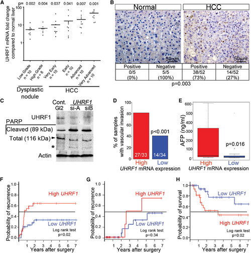
UHRF1 mRNA and Protein Are Overexpressed in HCC (A) UHRF1 detected by qRT-PCR in 18 preneoplastic lesions and 40 HCCs from hepatitis C virus (HCV)-infected patients compared to expression in nine normal livers. Horizontal line indicates median. (B) Immunohistochemistry for UHRF1 protein (brown) was evaluated in 52 of the same HCCs examined in (A) plus five normal liver samples. Fisher’s exact test was used to calculate p value. Seventy-one of the HCV-associated HCCs analyzed by qPCR were grouped into high (n = 35) and low (n = 36) UHRF1-expressing tumors based on the median log2-fold change of 3.64. (C) HepG2 cells transfected with control siRNA (GL2) or two different siRNAs targeting UHRF1 described in Tien et al. (2011) were blotted for UHRF1 and cleaved and total PARP (arrow indicates full length; * indicates cleaved protein). (D–H) Vascular invasion (33 high and 34 low tumors; four missing values) (D), serum AFP (29 UHRF1-high and 29 low tumors; 13 missing values) (E), early (<2 years) (F) and late (>2 years; 32 UHRF1-high and 35 low tumors; four missing values) (G) tumor recurrence, and overall survival after surgery (32 high and 35 low tumors; four missing values) (H) were stratified according to UHRF1 expression. Continuous and categorical variables were assessed by Wilcoxon rank-sum test and Fisher’s exact test, respectively. Clinical outcome difference was evaluated by log rank test. In box and whisker plots, boxes represent the 75th and 25th percentiles, the whiskers represent the most extreme data points within interquartile range 3 1.5, and the horizontal bar represents the median. |
Reprinted from Cancer Cell, 25(2), Mudbhary, R., Hoshida, Y., Chernyavskaya, Y., Jacob, V., Villanueva, A., Fiel, M.I., Chen, X., Kojima, K., Thung, S., Bronson, R.T., Lachenmayer, A., Revill, K., Alsinet, C., Sachidanandam, R., Desai, A., SenBanerjee, S., Ukomadu, C., Llovet, J.M., and Sadler, K.C., UHRF1 overexpression drives DNA hypomethylation and hepatocellular carcinoma, 196-209, Copyright (2014) with permission from Elsevier. Full text @ Cancer Cell