
Fig. S4
- ID
- ZDB-FIG-140527-66
- Publication
- Kubo et al., 2014 - Functional architecture of an optic flow-responsive area that drives horizontal eye movements in zebrafish
- Other Figures
- All Figure Page
- Back to All Figure Page
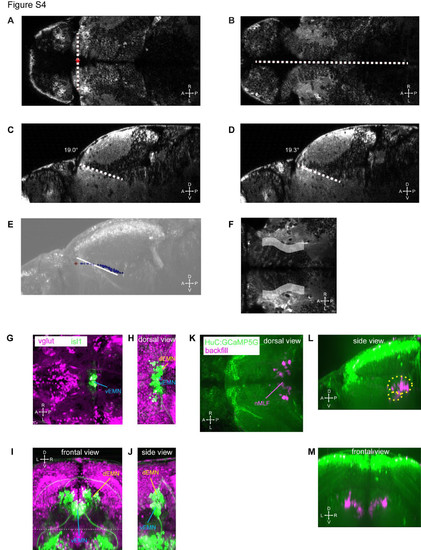
Registration procedure and landmarks (related to Figure 2). (A, B) Dorsal view of a HuC:GCaMP5G larva. The xy-position of the origin (red asterisk) is defined by the intersection of a line connecting the anterior tips of the AF9 containing neuropil (A) and the midline (B). The dorsoventral position of the origin is defined by moving dorsally in the z-stack until the anterior tips of the AF9 containing neuropil are not visible anymore (A). (C, D) In sagittal view, lines are drawn along the dorsal border of the AF9 containing neuropil in the left (C) and right (D) hemispheres to measure the pitch angle of the animal relative to the imaging plane. (E) The precise position of the dorsal edge of the AF9 containing neuropil (blue points) and the origin (red asterisk) are overlaid on a projected sagittal view. In addition, the registration line from (C,D) is overlaid (white) to show that the course of the border of the neuropil at particular medio-lateral levels (C,D) does not capture the overall curvy shape that is evident when measuring the medio-lateral center points of the dorsal edge of the neuropil (blue points). (F) The schematic neuropil landmark (gray, z-projected) is overlaid on a single optical slice located 40 μm below the posterior commissure (dorsal view). Note that the schematic shape does not match the medio-lateral extent of the neuropil, since the schematic shape only corresponds to the dorsal border of the AF9 containing neuropil and the more dorsally located neuropil (see e.g. Figure S4B) is located further medially. (G-J) The dorsal extraocular motor neurons (dEMN) and ventral extraocular motor neurons (vEMN) were imaged in vglut:dsRed; isl1:GFP transgenic larvae. (G) Optical slice at the dorso-ventral center of the vEMN (dorsal view). (H-J) Dorsal, frontal and sagittal images were projected along the extent of dEMN and vEMN. Image volumes dorsal to the dashed line in (I) are projected in (H). (K-M) Projected images of one of the backfilled HuC:GCaMP5G larvae used for measuring the position of the nucleus of the medial longitudinal fasciculus (nMLF). The approximate shape of the nMLF landmark used in our registered 3D maps is indicated in (L) by yellow, dashed ellipsoids. |