
Fig. 4
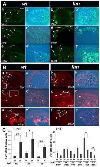
fan mutants exhibit increased apoptosis and reduced cell proliferation. (A) TUNEL Staining. Wild type embryos at 16 hpf (a, a2), 24 hpf (c, c2) and 48 hpf (e, e2) exhibited few apoptotic cells at all stages (arrows). In contrast, age matched fan mutant embryos exhibited increased levels of apoptotic cells at all stages examined (b, b2, d, d2, f, f2, arrows). Abbreviations: e, eye; n, neural tissue; pa, pharyngeal arches; y, yolk). Scale bar = 100 µm. (B) pH3 Immunofluorescent (IF) histochemistry. At 16 hpf, 24 hpf and 48 hpf, wild type embryos exhibit discrete pH3 expression in proliferating cells of the eye (e), neural tissues (n), and pharyngeal arches (pa) (a, a2, c, c2, e, e2). In contrast, age matched fan mutants exhibited reduced pH3 positive cell proliferation at all stages examined (b, b2, d, d2, f, f2, arrows). (Abbreviations: y, yolk). Scale bar = 100 µm. For both (A) and (B), fluorescent (a–f) and bright field plus fluorescent (a2–f2) images are shown. (C) Quantification of TUNEL and pH3 IF. For TUNEL, all apoptotic cells in each panel were counted for comparison between age matched wild type and fan mutant embryos. For pH3 IF, red fluorescent cells were counted in boxed areas as indicated. These results showed that fan mutants exhibited significantly increased apoptosis at all stages examined and significantly reduced cell proliferation at 48 hpf. At least 3 sectioned embryos were examined for each genotype at each developmental stage. Statistical analyses were performed using Student′s t-test. (p = >0.01). |
| Fish: | |
|---|---|
| Observed In: | |
| Stage Range: | 14-19 somites to Long-pec |