
Fig. 5
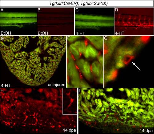
Regenerated coronary endothelium derives from preexisting endothelial cells during zebrafish heart regeneration. (A–D) Permanent labeling of endothelial cells during zebrafish embryogenesis. Tails of 96-hpf Tg(kdrl:CreER); Tg(ubi:Switch) embryos treated between 24 and 48 hpf with EtOH (A and B) or 4-HT (C and D) and imaged in the green (A and C) and red (B and D) channels. All 4-HT–treated embryos examined expressed mCherry in endothelial cells (n > 100), whereas EtOH-treated embryos failed to report red fluorescence (n > 100). (E–G) The adult endocardium and coronary endothelium displayed widespread and permanent mCherry labeling following 4-HT treatment during embryonic stages (n = 3/3). Representative cardiac section (E) and close up views of mCherry-labeled endocardium (F) and coronary endothelium (G). (H and I) Representative cardiac section from a regenerating Tg(kdrl:CreER); Tg(ubi:Switch) animal with mCherry-labeled endocardial and endothelial cells at 14 dpa. Numerous mCherry+ endothelial tubes (Inset) populated the regenerating myocardium. (n = 3/3.) |