
FIGURE
Fig. 2
- ID
- ZDB-FIG-140425-23
- Publication
- Rengarajan et al., 2014 - Endocytosis of Fgf8 is a double-stage process and regulates spreading and signaling
- Other Figures
- All Figure Page
- Back to All Figure Page
Fig. 2
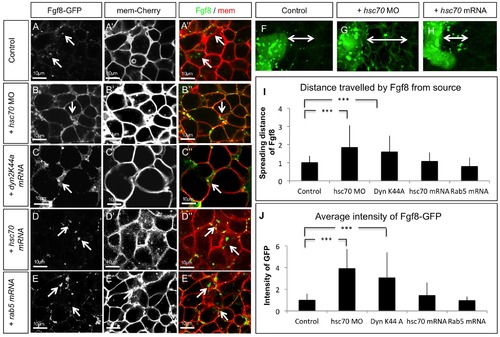
Fgf8 uptake is regulated by Hsc70. Confocal analysis of live embryos at 50% epiboly stage at the animal pole. Fgf8-GFP DNA was injected along with the red membrane marker mCherry at the one cell stage to determine subcellular localization of Fgf8 in embryos co-injected with indicated constructs (A–E′′). The range of Fgf8 propagation in the receiving tissue was analyzed using confocal microscopy (F–H) and the distance spread by Fgf8 was quantified (I). Quantification of the average intensity of Fgf8-GFP is demonstrated in embryos (J). For quantification 7 different embryos were used for each experiment. |
Expression Data
Expression Detail
Antibody Labeling
Phenotype Data
Phenotype Detail
Acknowledgments
This image is the copyrighted work of the attributed author or publisher, and
ZFIN has permission only to display this image to its users.
Additional permissions should be obtained from the applicable author or publisher of the image.
Full text @ PLoS One