
Fig. 6
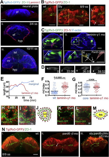
Laminin1 and Pard6γβ Are Required for Eye Morphogenesis Progression (A) Eye field and optic vesicles of fixed Tg(rx3:gfp) embryos immunostained against ZO-1 (green), Laminin1 (red), and GFP (blue). (B) Tg(rx3:gfp) and Tg(rx3:gfp)/sly optic vesicles immunostained against GFP (red) and ZO-1 (green). White arrows point to ZO-1 mis-localizing basally within the optic vesicles. (C) Transplants of Tg(rx3:gfp) expressing cells into nontransgenic hosts are shown: wild-type cells into wild-type embryos (Ci) and lamininγ1 morphant cells into lamininγ1 morphant embryos (Cii), immunostained against GFP (green), ZO-1 (red), and F-actin (blue), showing the disrupted epithelial organization (Cii) and the aberrant AB polarity axis (Ciii) in lamininγ1 morphants. (D) Time-lapse movie of a lamininγ1 morphant marginal cell expressing Pard3-GFP. (E) Changes in cell length over time of a wild-type (blue) and lamininγ1 morphant (red) marginal cell transplanted into wild-type and morphant hosts, respectively. (F) Measurement of cell rdn index of 6–7 ss eye field cells in a wild-type (blue) and lamininγ1 morphant embryo (red); lamininγ1 morphant eye field cells are rounder than wild-type eye cells (wild-type, 0.34 ± 0.18; lamininγ1 MO, 0.58 ± 0.18, mean ± SD). Black bar indicates mean (Mann-Whitney U test). WT, n = 83 cells in five embryos, Lamininγ1 mo n = 66 cells in four embryos, p < 0.0001. All eye field cells (marginal and core) have been included in the measurement. (G) lamininγ1 morphant eye cells (presumptive core and marginal in red) are similar in their rdn index to core wild-type cells (blue). Black bar indicates mean (Mann-Whitney U test). WT core cells, n = 53 core cells in five embryos; Lamininγ1 morphant, n = 66 cells in four embryos, p = 0.5436. (H, I, K, and L) Images showing apical polarity organization of cells (labeled by Pard3-GFP, green) in the optic vesicles of embryos in which Laminin1-coated (H and I) or BSA-coated (K and L) beads were implanted in the center of the eye field. See also Movie S9. BF, bright field. (J and M) Schematics illustrating the organization of cells around beads in the eyes from (H) and (K). (N) Tg(rx3:GFP) (Ni), Tg(rx3:GFP);pard6γβMo (Nii), and Tg(rx3:GFP);sly;pard6γβMo (Niii) embryos stained as detailed in the figure. The arrow in (Nii) points at defective polarization in pard6γβ morphants. White lines in (C), (H), and (I) outline the margins of the eye field. Scale bars, 27 µm (A–D and N). See also Figure S4 and Movie S9. |
Reprinted from Developmental Cell, 27(3), Ivanovitch, K., Cavodeassi, F., and Wilson, S.W., Precocious acquisition of neuroepithelial character in the eye field underlies the onset of eye morphogenesis, 293-305, Copyright (2013) with permission from Elsevier. Full text @ Dev. Cell