
Fig. 3
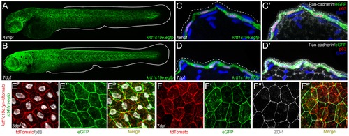
Characterization of krtt1c19e :egfp transgenic larvae. A-D′: Confocal images of eGFP expression in germline krtt1c19e:egfp transgenic larvae at 48 hpf (A, C-C′) and 7 dpf (B, D-D′). A-B: Lateral overviews of transgenic larvae indicating the krtt1c19e promoter drives eGFP expression in the epidermis of zebrafish larvae at all locations except the fins (extent of medial fins outlined by white line). C-D′: Immunofluorescent labelling of transgenic larvae cryosections demonstrates co-expression of eGFP (green; C-D′) and ΔNp63 (red; C-D′) in basal keratinocytes. Counterstaining with DAPI (blue; C-D′) and Pan-cadherin (white; C′, D′) highlights the eGFP negative region overlying EVL (demarcated by dashed lines; C′, D′). E-F′′′: Confocal images of the epidermis of germline krtt1c19e:lyn-tdtomato; krt4:lyn-egfp double transgenic larvae at 72 hpf (E-E′′) and 7 dpf (F-F′′′) immunofluorescently stained for eGFP (green; E′-E′′, F′, F′′′), tdTomato (red; E, E′′, F, F′′′), ΔNp63 (white; E, E′′) and ZO-1 (white; F′′-F′′′). The expression of membrane bound tdTomato delineates the ΔNp63 positive basal keratinocytes from the eGFP expressing ZO-1 positive EVL cells. |
| Genes: | |
|---|---|
| Antibodies: | |
| Fish: | |
| Anatomical Terms: | |
| Stage Range: | Long-pec to Days 7-13 |