
Fig. S2
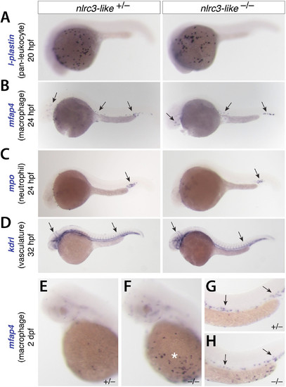
Early formation of primitive macrophages, neutrophils, and the vasculature appear normal in nlrc3-like–/– mutants, Related to Figure 2. The images show embryos analyzed by whole mount in situ hybridization. (A) At 20 hpf there was no apparent difference in expression in l-plastin, a marker of all leukocytes. (B) At 24 hpf, mfap4 expression indicated normal formation and migration of primitive macrophages into the embryo proper in mutants and siblings (arrows). (C) At 24 hpf, mpo expression showed no difference (arrow) between mutants and siblings. (D) Vasculature formed normally in the mutants, based on the expression pattern of the endothelial marker kdrl at 32 hpf (arrows). (E–H) mfap4 expression at 2 dpf shows the abundance of macrophages in the periphery of nlrc3-like mutants. Compared with heterozygous siblings (E), mutants (F) have abnormal aggregations of macrophages in the yolk sac (asterisk), but similar pattern of macrophages in the caudal vein (G–H, arrows). All panels show lateral views, with anterior to the left and dorsal to the top. |