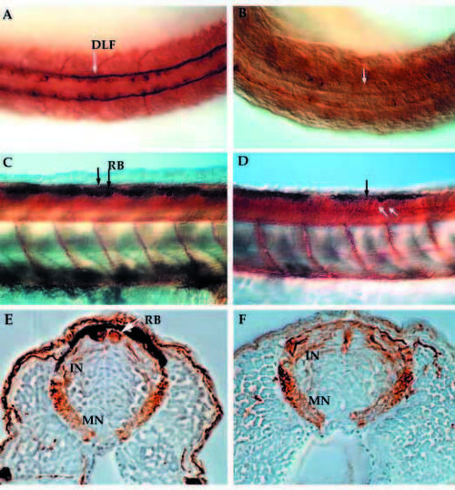
Anti-acetylated α-tubulin immunostained whole-mount embryos at 24 hpf (A,B) and 48 hpf (C-F) reveal the neuronal pattern in nrd. Dorsal view of the neuronal pattern of a 24 hpf wild-type embryo (A) shows large RB neurons (arrow; large brown cells) as well as a distinct DLF (two parallel tracts of axons). In nrd (B), the embryos have very little dorsal expression of a-tubulin, except in a few scattered commissural neurons (dark brown stained cells). At 48 hpf, the differentiation of many neuronal cell types including large RB neurons can be seen in wild-type embryos (C, arrows and lateral view). In nrd (D), other neurons such as commissural neurons appear to develop normally (white arrows), but RB neurons do not form (black arrow point to area where RBs should form). (E,F) Transverse sections of embryos in C and D (embryos were embedded in plastic and sectioned at a thickness of 3 mm). In wild-type embryos, RBs are large neurons at the dorsal most aspect of the neural tube (E), while nrd embryos lack RBs. RB, Rohon Beard cell; DLF, dorsal longitudinal fascicle; In, interneuron; MN, motorneuron.
|
