
Fig. 6
- ID
- ZDB-FIG-140313-9
- Publication
- Warga et al., 1999 - Origin and development of the zebrafish endoderm
- Other Figures
- All Figure Page
- Back to All Figure Page
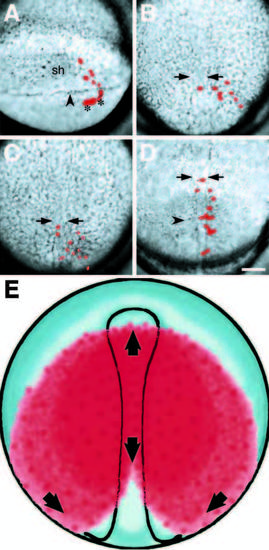
Endodermal precursors are stereotypically distributed in the late gastrula. (A-D) Illustrate the convergence of endodermal precursors towards the dorsal midline from selected composite frames of a time-lapse video. Labeled cells are pseudocolored red. (A) The clone at shield stage, consisting of six deep cells and four enveloping layer cells (asterisks), to the right of the embryonic shield (sh). The labeled deep cells have already involuted deep into the hypoblast; arrowhead, blastoderm margin. (B) By 80% epiboly, labeled cells have begun to converge towards the embryonic axis whose boundaries are indicated with arrows for this and subsequent panels. (C) By 100% epiboly, labeled cells have begun to spread towards the animal pole. (D) At the 4-somite stage, the labeled cells now extend anterior-posteriorly along the axis. The more anterior cells were in the prechordal plate and gave rise to pharynx, a common prechordal plate derivative in our data set, the more posterior cells gave rise to liver, swimbladder and stomach; arrowhead, first somite furrow. (E) Illustrates the spatial distribution of endodermal precursors in the late gastrula based on our data. Red indicates where cells of the endoderm were observed, and arrows indicate directions cells move to meet at the dorsal midline and spread antero-posteriorly. Scale bar: 100 μm. |