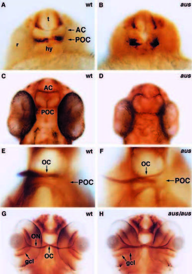
Commissure formation is delayed and perturbed in aus mutant embryos. Frontal/ventral views of whole-mount embryos stained with an antibody against acetylated tubulin focussed at the level of the anterior and postoptic commissures. (A,B) Prim-5 stage embryos. The anterior commissure and postoptic commissure are not formed in the aus mutant embryo. (C,D) Prim-25 stage embryos. By this stage, some axons have crossed the midline in both commissures in the aus mutant embryo. (E,F) Protruding-mouth stage embryos. The postoptic commissure and optic chiasm are defasciculated and slightly disorganised in the aus mutant embryo. (G,H) Protrudingmouth stage embryos. The optic axons are less tightly fasciculated as they exit the eye of the putative homozygous aus mutant embryo as compared to the wild-type sibling. The failure of the choroid fissure to fully close (coloboma) results in the retinal ganglion cells protruding towards the midline. Abbreviations: AC, anterior commissure; gcl, ganglion cell layer; hy, hypothalamus; OC, optic chiasm; ON, optic nerve; POC, postoptic commissure; r, retina; t, telencephalon. Scale bar: (A-D,G,H) 25 μm; (E,F) 10 μm.
|
