
Fig. 4
- ID
- ZDB-FIG-140303-37
- Publication
- Moens et al., 1996 - valentino, a zebrafish gene required for normal hindbrain segmentation
- Other Figures
- All Figure Page
- Back to All Figure Page
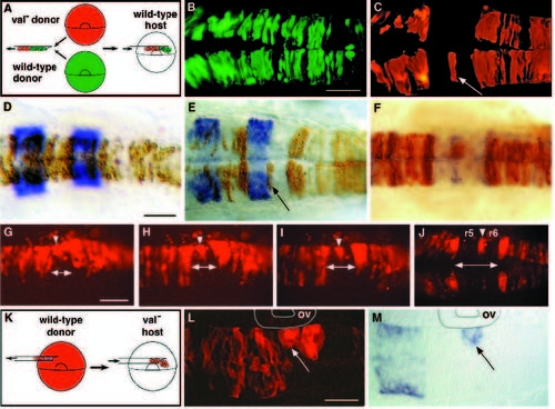
Mosaic analysis demonstrates that valentino is required for cells to contribute to either r5 or r6. (A) Schematic diagram of the experimental approach used for B-J. Rhodaminelabeled cells from a val- donor and fluoresceinlabeled cells from a wildtype donor were transplanted into the same unlabeled wild-type host at the early gastrula stage (see Materials and Methods). The distribution of labeled cells was then observed at 24 h of development. (B,C) Confocal images taken in dorsal view showing the distribution of wild-type (B) and val- cells (C) in the hindbrain region of the same wild-type host. val- cells are specifically excluded from a sharply defined region of the hindbrain, but are otherwise able to contribute to the entire brain and spinal cord. A single val- cell or small cluster of val- cells is frequently observed at the center of this region (arrow in C). (D-F) krox20 staining (D,E) and mariposa staining (F) of transplant recipients. Brown cells are donor-derived (see Materials and Methods). (D) The distribution of wild-type cells in the hindbrain of a wild-type host. (E,F) The distribution of val- cells in a wild-type host, showing that mutant cells are specifically excluded from r5 and r6. The embryo shown in E is the same embryo as is shown in B and C, and the cell noted in C is observed to lie at the r5-6 boundary (arrow in E). In 31 out of 39 genetically mosaic embryos analyzed in which transplanted cells had contributed to the hindbrain, labelled val- cells were excluded from r5 and r6 although not necessarily from the boundary between them. In the remaining 8 mosaic embryos, val- cells were observed in r5 and/or r6, but these cells were located ventrally, either in or very near the floor plate (not shown). In contrast, 94 out of 100 control embryos into which labeled wild-type cells had been transplanted had labeled cells distributed throughout the hindbrain, including r5 and r6. The remaining 6 control embryos had relatively few transplanted cells and these tended to be localized to rhombomere boundaries. (G-J) A series of live images of rhodamine-labeled val- cells transplanted into a single wild-type host, shown in dorsal view. The time-points shown are 7 somites (12 h; G), 10 somites (14 h; H), 14 somites (16 h; I) and 24 h (J). As early as the 7-somite stage, val- cells begin to be excluded from the presumptive r5 and r6 of a wild-type host (indicated by a double arrow at the midline), with the exception of a few cells that ultimately lie at the r5-6 boundary (arrowheads). (K) Schematic diagram of the experimental approach used for L and M. Rhodamine-labeled cells from a wild-type donor were transplanted into a val- host embryo at the early gastrula stage. (L,M) Fluorescent and transmitted light images of the same horizontal section of a mutant host embryo. Anterior is to the left. Donor-derived cells in this experiment are fluorescently labelled. (L) Wild-type cells in a val- host form abnormal, unilateral clusters of rounded cells in rX, which is adjacent to the otic vesicle (ov), while they otherwise contribute normally to the brain and spinal cord. (M) The more anterior of these clusters, which lies at the r4-rX boundary, autonomously expresses krox20 although the surrounding mutant cells have failed to acquire r5 identity (arrow in L and M). Scale bars: B,C, 100 μm; D-F, 50 μm; G-J, 100 μm; L,M, 50 μm. |