
Fig. 9
- ID
- ZDB-FIG-140303-21
- Publication
- Concordet et al., 1996 - Spatial regulation of a zebrafish patched homologue reflects the roles of sonic hedgehog and protein kinase A in neural tube and somite patterning
- Other Figures
- All Figure Page
- Back to All Figure Page
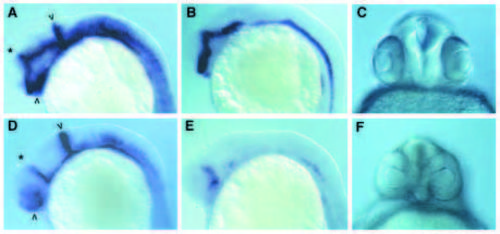
Suppression of midline signalling by cPKA activity. (A-C) normal (uninjected embryos) (D-F) embryos injected with cPKA RNA at the 2- to 4-cell stage. (A,D) were hybridised with probes for both pax[b] and ptc1. In D, most of the signal derives from the pax[b] probe including the stripe at the midbrain/hindbrain boundary (arrowhead) and the reduced staining in the optic stalks (arrowhead). The asterisk indicates the stripe of ptc1 expression in the diencephalon - note that in the injected embryo this extends further ventrally compared to the wild-type embryo, indicating a dorsalisation of the brain. (B,E) Hybridised with a probe for nk2.2. Note the marked loss of expression following injection of the cPKA mRNA. (C,F) Frontal views of the heads of 27- hour-old embryos, showing the partial fusion of the eyes in the cPKA-injected animal. |