|
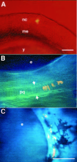
chn neural crest cells do not differentiate as cartilage in the arches. (A) 12 hour, lateral view. A single neural crest cell, labeled with tetramethylrhodamine-dextran is shown after injection. (B,C) 72 hour, lateral view. Computer-combined bright field and fluorescence images. Clones in the mandibular arch in living wild-type (B) and chn mutant (C) embryos, derived from cells located in the largely chondrogenic region of the fate map at early segmentation stages (12-13 hours; Table 1). (B) In the wild type, the clone has contributed several cartilage cells to the palatoquadrate (between arrows). (C) Neural crest cells that migrate into the mandibular arch in chn, remain mesenchymal and never form cartilage although surrounding melanocytes, derived from separate unlabeled neural crest lineages develop normally (Schilling and Kimmel, 1994). Abbreviations: e, eye; me, paraxial mesoderm; nc, neural crest; pq, palatoquadrate; y, yolk. Scale bars, 100 μm.
|