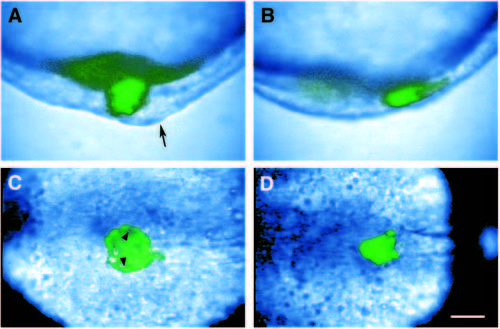
Caged fluorescein labeling of the forerunner population in wild-type and ntl mutant embryos. Composite video images of caged fluorescein labeling in wild-type (A,C) and ntl mutant (B, D) embryos. The forerunner cell population was labeled by uncaging during the early gastrula period (5.5-6.5h). Fluorescein-labeled cells are colored green. (A,B) Side views of the tailbud at the bud stage. Dorsal is to the right. In wild-type embryos, there is labeling in a cluster of cells located deep to the chordoneural hinge (arrow), the larger tissue mass on the dorsoanterior side of the tailbud. In ntl mutant embryos at the bud stage, the forerunner cell cluster appears to be adjacent to the chordoneural hinge, and the ventral side of the tailbud is proportionately larger. (C,D) Dorsal views of the tailbud at the 7-somite stage. The axial mesoderm extends anteriorly to the right. (C) By this stage, Kuppfer’s vesicle is obvious in the tailbud of wild-type embryos, arrowheads point to the inside wall of the vesicle. Cells lining Kupffer’s vesicle are specifically labeled. (D) In contrast, fluoresceinlabeled cells in ntl mutant embryos remain in a tight cluster. Scale bar, 50 μm in A,B; 72 μm in C,D.
|
