
FIGURE
Fig. 3
- ID
- ZDB-FIG-140226-18
- Publication
- Delaurier et al., 2014 - Role of mef2ca in developmental buffering of the zebrafish larval hyoid dermal skeleton
- Other Figures
- All Figure Page
- Back to All Figure Page
Fig. 3
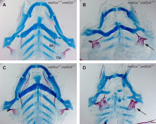
mef2cb may not function in OpBR development, but functions partially redundantly with mef2ca in cartilage development. Ventral views of dissected flat mounts at 6 dpf, stained with Alcian Blue and Alizarin Red. (A and C) The skeletons (bone, red; cartilage, blue) appear phenotypically normal. (B and C) The OpBR bones show the expansion phenotype (arrows), the cartilages are disrupted, substantially more so in (D) than (B). Scale bar 50 μm. |
Expression Data
Expression Detail
Antibody Labeling
Phenotype Data
| Fish: | |
|---|---|
| Observed In: | |
| Stage: | Day 6 |
Phenotype Detail
Acknowledgments
This image is the copyrighted work of the attributed author or publisher, and
ZFIN has permission only to display this image to its users.
Additional permissions should be obtained from the applicable author or publisher of the image.
Reprinted from Developmental Biology, 385(2), Delaurier, A., Huycke, T.R., Nichols, J.T., Swartz, M.E., Larsen, A., Walker, C., Dowd, J., Pan, L., Moens, C.B., and Kimmel, C.B., Role of mef2ca in developmental buffering of the zebrafish larval hyoid dermal skeleton, 189-99, Copyright (2014) with permission from Elsevier. Full text @ Dev. Biol.