
Fig. 3
- ID
- ZDB-FIG-140214-22
- Publication
- Begemann et al., 2000 - Developmental regulation of Tbx5 in zebrafish embryogenesis
- Other Figures
- All Figure Page
- Back to All Figure Page
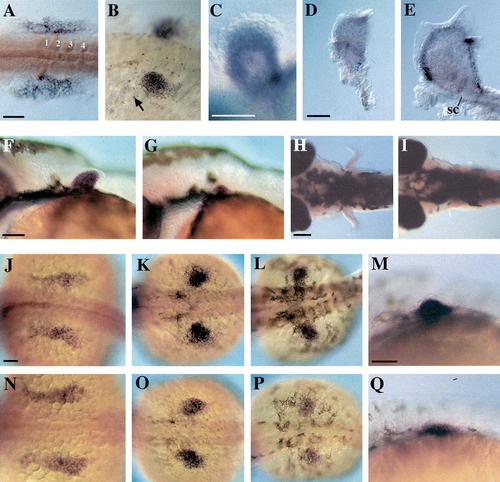
tbx5.1 expression in pectoral fin buds in wild type and mutant zebrafish. (A-E) tbx5.1 expression during wild type pectoral fin development. (A) 18-somite stage. Somites 1-4 are indicated. (B) 32 hpf. Arrow points to tbx5.1 expressing cells anterior to and outside of the pectoral fin bud. (C) 48 hpf. Expression is restricted to the distal part of the fin bud, which forms the fin endoskeleton proper. (D) 3 day embryo. The band of darker staining in the proximal mesenchyme is due to melanocytes that cover the epidermis at the ventral base of the fin bud. (E) 5 day embryo. Note the absence of tbx5.1 expression in the ectoderm of the apical fold and in the scapulocoracoid (sc). (F-I) tbx5.1 is expressed in dak mutant fin buds. (F) 36 hpf wild type and (G) daktw25 pectoral fin buds. (H, I) At 5 days, daktw25 mutants (I) lack differentiated pectoral fins. (J-Q) Maintenance of tbx5.1 in wild type (J-M) and syut4 mutant fin buds (N-Q). Expression at the 22-somite stage (J, N), 32 hpf (K, O), 36 hpf (L, M, P, Q). Anterior is to the left, (C-G, M, Q) lateral views with dorsal on top, (A, H-L, N-P) dorsal views. Scale bars, 100 µm. |
| Gene: | |
|---|---|
| Fish: | |
| Anatomical Terms: | |
| Stage Range: | 14-19 somites to Day 5 |
| Fish: | |
|---|---|
| Observed In: | |
| Stage Range: | Prim-25 to Day 5 |
Reprinted from Mechanisms of Development, 90(2), Begemann, G. and Ingham, P.W., Developmental regulation of Tbx5 in zebrafish embryogenesis, 299-304, Copyright (2000) with permission from Elsevier. Full text @ Mech. Dev.