
Fig. 3
- ID
- ZDB-FIG-140130-34
- Publication
- Fleming et al., 2013 - Functional characterisation of the maturation of the blood-brain barrier in larval zebrafish
- Other Figures
- All Figure Page
- Back to All Figure Page
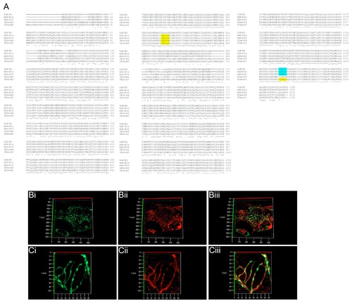
Sequence comparison and expression of zebrafish ABCB1/4/5 homologues. A) Sequence alignment of human and mouse ABCB1 protein sequences with zebrafish Abcb4 and Abcb5. The zebrafish proteins show a high level of identity with the mouse and human ABCB1, ABCB4 and ABCB5 proteins. An antibody recognising the peptide sequences VQAALD (yellow) and VQEALD (blue) (Covance) was selected for use in zebrafish as the similarity of these peptides was conserved. B (low magnification) and C) (high magnification) 3D projections of optically sectioned wholemount Tg(fli1a:EGFP)y1 larvae stained with the anti-VQAALD antibody. Positive staining is observed in the vascular endothelium of the CNS at 8 d.p.f , but not at earlier timepoints (see Figure S2). High magnification images (Ci-iii) demonstrate the co-localisation of ABCB1/4/5 antibody staining (red) on cerebral vessels (green). Bi and Ci – GFP channel – maximum intensity projection of the cerebral vasculature of Tg(fli1a:EGFP)y1 transgenic larvae; Bii and Cii – Alexa 568 labelled antibody staining with ABCB1/4/5 antibody; Biii and Ciii – overlay. |
| Antibody: | |
|---|---|
| Fish: | |
| Anatomical Terms: | |
| Stage: | Days 7-13 |