
Fig. 4
- ID
- ZDB-FIG-140130-31
- Publication
- Kochhan et al., 2013 - Blood Flow Changes Coincide with Cellular Rearrangements during Blood Vessel Pruning in Zebrafish Embryos
- Other Figures
- All Figure Page
- Back to All Figure Page
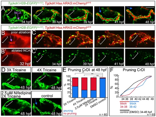
Pruning of the CrDI is flow dependent. Lateral views, anterior to the left, dorsal to the top. (A) Still images of time-lapse of Tg(kdrl:EGFP)s843; Tg(gata1a:DsRed)sd2 from 34–48 hpf. White brackets indicate dorsal CrDI, purple arrow indicates NCA. Note reduction of blood flow in dorsal CrDI (39 hpf time point) prior to regression (blue arrows at 41 and 48 hpf time points). (B) Laser ablation of NCA at 32 hpf (purple arrow), compare to (B′). (C) Time-lapse imaging from 34–48 hpf of Tg(kdrl:H2B-EGFP)mu122; Tg(kdrl:Hsa.HRAS-mCherry)s916 after NCA ablation and Tg(kdrl:H2B-EGFP)mu122 only (C′). Note persistent CrDI (white brackets). (D) Blocking of heartbeat by indicated drug treatments from 30–48 hpf and imaging at 48 hpf. (E) Quantification of drug treatments shown in D. (F) Influence of 4 hr block of heartbeat followed by drug wash out on CrDI regression at indicated time points. Scale bar = 50 μm. |
| Genes: | |
|---|---|
| Fish: | |
| Anatomical Terms: | |
| Stage Range: | Prim-15 to Long-pec |