
Fig. 4
- ID
- ZDB-FIG-140122-42
- Publication
- Kang et al., 2013 - Local dkk1 crosstalk from breeding ornaments impedes regeneration of injured male zebrafish fins
- Other Figures
- All Figure Page
- Back to All Figure Page
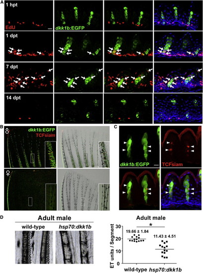
Male Pectoral Fin ETs Undergo Vigorous Renewal (A) Section images of male dkk1b:EGFP pectoral fin ETs at 1 hr, 1 day, 7 days, or 14 days after EdU injection. Arrows indicate EdU+dkk1b:EGFP-expressing cells. Green indicates enhanced green fluorescent protein (EGFP) immunofluorescence, red indicates EdU immunofluorescence, and blue indicates DAPI. Scale bar, 10 μm. (B) Whole-mount images of TCFsiam; dkk1b:EGFP pectoral fins. Males expressed both reporters in ETs. (C) Section image of maleTCFsiam; dkk1b:EGFP pectoral fin ETs. Arrowheads indicate TCFsiam+ cells. Cuticle shows red autofluorescence in these images. Scale bar, 10 μm. (D) Ubiquitous overexpression of dkk1b decreased the number of ETs in male pectoral fins and reduced their definition. Data are shown as mean ± SD. p < 0.001 by two-tailed Student’s t test. |
Reprinted from Developmental Cell, 27(1), Kang, J., Nachtrab, G., and Poss, K.D., Local dkk1 crosstalk from breeding ornaments impedes regeneration of injured male zebrafish fins, 19-31, Copyright (2013) with permission from Elsevier. Full text @ Dev. Cell