
Fig. S4
- ID
- ZDB-FIG-140114-5
- Publication
- de Oliveira-Carlos et al., 2013 - Notch receptor expression in neurogenic regions of the adult zebrafish brain
- Other Figures
- All Figure Page
- Back to All Figure Page
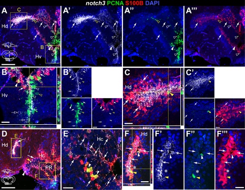
notch3 expression in glia and proliferating cells of the adult zebrafish hypothalamus. Confocal images showing localization of notch3 mRNA by FISH (white), radial glia labelled with S100β (red), and PCNA + proliferating cells (green). Cross-sections at the indicated levels through the diencephalon; hypothalamic area shown in the micrographs is indicated in the cross-section schematics. A, B, notch3 is expressed in most PCNA+ /100β- cells (filled white arrowheads) and in PCNA +/S100β - cells (unfilled white arrowheads) of the Hv; unfilled yellow arrowheads indicate notch3 - /PCNA +/S100β cells. A–F, notch3 localizes with most S100β cells of Hd and Hc, PCNA + (filled white arrowheads) or PCNA (filled white arrows); filled yellow arrowhead indicates a notch3 -/PCNA +/S100β + cell. Notice the notch3 expression in the S100β + cellular processes in Hc (in E). Asterisk indicates a S100β group of cells in Hd that is negative for notch3. Abbreviations: Hc, caudal zone of the periventricular hypothalamus; Hd, dorsal zone of the periventricular hypothalamus; Hv, ventral zone of the periventricular hypothalamus: PR, posterior recess of the diencephalic ventricle. Scale bar = 100 μm in A and D, 20 μm in B, B , C, C , E, F, F . |