
FIGURE
Fig. 4
- ID
- ZDB-FIG-131218-57
- Publication
- Cavodeassi et al., 2013 - Eph/Ephrin signalling maintains eye field segregation from adjacent neural plate territories during forebrain morphogenesis
- Other Figures
- All Figure Page
- Back to All Figure Page
Fig. 4
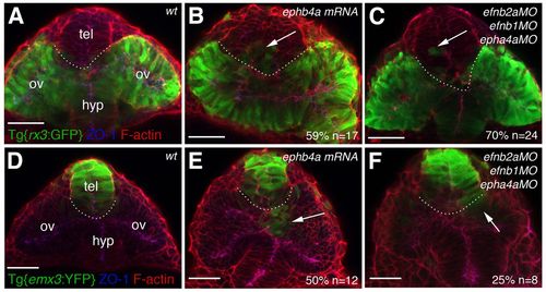
Manipulation of Eph/Ephrin activity leads to defective segregation of eye and telencephalic cells. Frontal views of the forebrain and eyes in Tg{rx3:GFP} (A-C) and Tg{emx3:YFP} (D-F) 10-12 ss wild-type, morphant or mRNA-injected zebrafish embryos immunostained to detect GFP (green), ZO-1 (blue) and F-actin (red). Arrows point at eye fated cells located in the telencephalic domain (B,C) or at telencephalic fated cells located in the optic vesicles (E,F). The dotted lines demarcate the transition between telencephalon and eye. ov, optic vesicle; tel, telencephalon; hyp, hypothalamus. Scale bars: 50 μm. |
Expression Data
| Genes: | |
|---|---|
| Antibody: | |
| Fish: | |
| Knockdown Reagents: | |
| Anatomical Terms: | |
| Stage: | 10-13 somites |
Expression Detail
Antibody Labeling
Phenotype Data
| Fish: | |
|---|---|
| Knockdown Reagents: | |
| Observed In: | |
| Stage: | 10-13 somites |
Phenotype Detail
Acknowledgments
This image is the copyrighted work of the attributed author or publisher, and
ZFIN has permission only to display this image to its users.
Additional permissions should be obtained from the applicable author or publisher of the image.
Full text @ Development