
Fig. 7
- ID
- ZDB-FIG-131218-11
- Publication
- Wang et al., 2013 - Fibronectin is deposited by injury-activated epicardial cells and is necessary for zebrafish heart regeneration
- Other Figures
- All Figure Page
- Back to All Figure Page
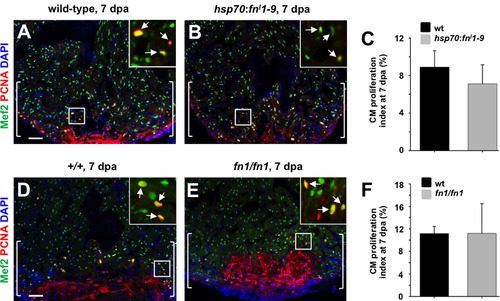
Evidence that Fibronectin does not directly regulate cardiomyocyte proliferation during regeneration. (A and B) Assessment of Mef2+PCNA+ cells (arrows) in hsp70:fnI1–9 (A) and clutchmate (B) 7 dpa ventricles. Insets, enlarged view of the rectangle. Brackets indicate injury site. (C) Quantification of cardiomyocyte proliferation in hsp70:fnI1–9 and clutchmate 7 dpa ventricles. For each group, 6 zebrafish were assessed. Student′s t-test, p=0.23. Mean±s.e.m. (D and E) Assessment of Mef2+PCNA+ cells (arrows) in wild-type (D) and homozygous fn1 mutant (E) 7 dpa ventricles. Brackets indicate injury site. (F) Quantification of cardiomyocyte proliferation in wild-type and homozygous fn1 mutant 7 dpa ventricles. For each group, 3–4 zebrafish were assessed. Student′s t-test, p=0.99. Mean±s.e.m. Scale bars: 50 μm. |
Reprinted from Developmental Biology, 382(2), Wang, J., Karra, R., Dickson, A.L., and Poss, K.D., Fibronectin is deposited by injury-activated epicardial cells and is necessary for zebrafish heart regeneration, 427-435, Copyright (2013) with permission from Elsevier. Full text @ Dev. Biol.