
Fig. S1
- ID
- ZDB-FIG-131211-8
- Publication
- Zhen et al., 2013 - Hemogenic endothelium specification and hematopoietic stem cell maintenance employ distinct Scl isoforms
- Other Figures
- All Figure Page
- Back to All Figure Page
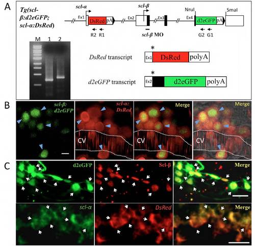
The transcription and expression of d2eGFP and DsRed recapitulate that of endogenous scl-β and scl-α. (A) 5′- RACE experiment to determine the transcription start sites of DsRed and d2eGFP. The position of respective 5′-RACE primer set is indicated (arrows; R1 and R2 for DsRed; G1 and G2 for d2eGFP). Line 1 and line 2 show the 5′-RACE PCR products of DsRed and d2eGFP. M, 1 kb DNA ladder. The sequence result of 59-RACE PCR products indicates that the DsRed transcript starts from the scl-α transcription initiation site and contains the non-coding exon 1, DsRed and SV40 poly(A) sequence; the d2eGFP transcript starts form the scl-&beta initiation site and contains the non-coding sequences of exon 2, Scl-β coding sequences of exon 2, 3 and part of exon 4 (black box), followed by the d2eGFP and SV40 poly(A) sequence. The asterisks indicate the translation initiation site of DsRed and d2eGFP, respectively. The d2eGFP protein is translated as a chimeric protein fused with the N-terminal 75 amino acids of Scl-β. (B) The expression of d2eGFP and DsRed in hematopoietic stem and progenitor cells in the caudal hematopoietic tissue (CHT) of 3 dpf Tg(scl- β:d2eGFP; scl-α:DsRed) larvae. D2eGFP is observed with nucleus restriction. Scale bar: 5 μm. CV, caudal vein. (C) Upper panel shows the double immunohistochemistry staining of d2eGFP and Scl-β (detected by Ab-Scl-C) in the anterior lateral plate mesoderm, where only Scl-β is expressed. Lower panel shows the double whole-mount in situ hybridization (WISH) of DsRed and scl-α (detected by scl-5′ probe) in the ICM of 22 hpf Tg(scl-β:d2eGFP;scl-α:DsRed) embryos. Arrows indicate the colocalized cells. Embryos are shown in lateral views with anterior to the left. Scale bars: 20 μm. |