
Fig. S8
- ID
- ZDB-FIG-131031-12
- Publication
- Suzuki et al., 2013 - Cone photoreceptor types in zebrafish are generated by symmetric terminal divisions of dedicated precursors
- Other Figures
- All Figure Page
- Back to All Figure Page
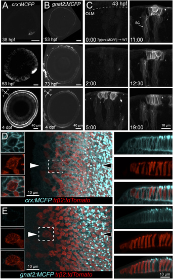
Time course of crx and gnat2 promoter activity in transient expression and stable transgenic lines. (A and B) FP expression driven by the crx or gnat2 promoter shown here in multiphoton image stacks of the eye at various ages. FP signal was first observed in the ventronasal patch at 38 hpf in Tg(crx:MCFP) animals. Subsequently, expression spread across the entire retina, localized to photoreceptors and bipolar cells. In contrast, FP expression driven by the gnat2 promoter was observed after 3 dpf, and expression was only observed in cones [also see Kennedy et al. (15)]. D, dorsal; N, nasal; T, temporal; V, ventral. (C) Cells from Tg(crx:MCFP) were transplanted into host embryos to facilitate tracking a small number of cells by time-lapse microscopy. Shown here is a 19-h duration time-lapse sequence (30-min intervals). MCFP expression is detected around 45 hpf and localized to cones and presumably dividing cone precursor cells. Fluorescence is also observed in differentiating bipolar cells (e.g., arrow). (D and E) To correlate the temporal expression of FP driven by the crx, gnat2, and trβ2 promoters, we crossed Tg(trβ2:tdTomato) with Tg(crx:MCFP) or Tg(gnat2:MCFP) animals. Confocal reconstructions of the region near the marginal zone. Note that MCFP signal is detected in dividing cells under the crx promoter but not under the gnat2 promoter. This suggests that, in the transgenic lines, crx-driven expression occurs at least at the stage of cone-precursor division, whereas gnat2-driven expression occurs in postmitotic cones. Orthogonal views shown represent views obtained along a line joining the two arrowheads. Nontrβ2:tdTomato-expressing cells also express FP under the crx or gnat2 promoters. The nasal side is toward the top of the images. |