
Fig. S4
- ID
- ZDB-FIG-131029-55
- Publication
- Fang et al., 2013 - Translational profiling of cardiomyocytes identifies an early Jak1/Stat3 injury response required for zebrafish heart regeneration
- Other Figures
- All Figure Page
- Back to All Figure Page
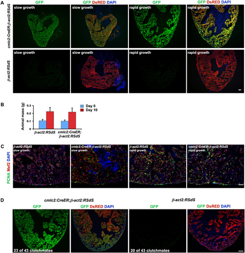
Inhibition of Stat3 in cardiomyocytes has no detectable effects during rapid animal growth. (A) Induction of dnStat3-GFP fluorescence in cardiomyocytes after 4-HT incubation in cmlc2:CreER;β-act2:RSdS or control β-act2:RSdS fish, during slow (normal) and accelerated growth conditions. (B) Control (β-act2:RSdS) and cmlc2:CreER;β-act2:RSdS animals each approximately doubled their mass after 10 d of rapid growth conditions. Data are mean ± SEM n = 12. (C) Representative images of cardiomyocyte proliferation using antibodies against PCNA and Mef2, indicating no detectable difference between control (β-act2:RSdS) and dnStat3-expressing fish (cmlc2:CreER; β-act2:RSdS) after 10 d of either slow or rapid growth. Quantified data are in Fig. 3D. (D) Control (β-act2:RSdS) and cmlc2:CreER;β-act2:RSdS embryos were incubated in 4-HT at 4 d postfertilization and allowed to grow to adulthood. Approximately half of the surviving animals from a clutch of 43 fish were cmlc2:CreER; β-act2:RSdS, indicated by myocardial dnStat3-GFP fluorescence. (Scale bars, 50 μm.) |