|
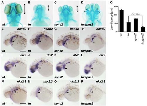
Lower jaw morphology. (A–D) Lower jaw morphology at 4dpf was visualized by Alcian Blue staining (ventral view). Anterior–posterior distances of the ventral pharyngeal arch (*) is indicated by the length of the double-headed arrows. The dorsal pharyngeal structure is identified by the crosses (+). (E–P) Whole-mount in situ hybridization using hand2, dlx2 and nkx2.3 RNA probes. The anteroventral position of these markers is marked by the arrowheads. All images show lateral views at 30hpf. Genotyping was performed by genomic sequencing after taking pictures. wt (A,E,I,M), fn mutant (B,F,J,N), spns2 mutant (C,G,K,O), and fn;spns2 double mutant (D,H,L,P). Scale bars: 200μm. (Q) Average anterior–posterior distances of the ventral pharyngeal arch from multiple experiments; error bars represent standard deviations.
|