
FIGURE
Fig. S3
Fig. S3
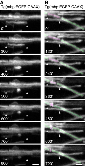
Time-lapse analyses of myelin sheath retractions Relates to Figure 3 and Movies S4 and S5. (A+B) Images from time-lapse analyses of Tg(mbp:EGFP-CAAX) animals showing myelin sheath retractions. In one example (A) the myelin sheath is retracted from an axon without myelin sheaths nearby, whereas in (B) the myelin sheath is being retracted from an axon that retains neighbouring myelin sheaths. Scale bar=5μm. |
Expression Data
Expression Detail
Antibody Labeling
Phenotype Data
Phenotype Detail
Acknowledgments
This image is the copyrighted work of the attributed author or publisher, and
ZFIN has permission only to display this image to its users.
Additional permissions should be obtained from the applicable author or publisher of the image.
Reprinted from Developmental Cell, 25(6), Czopka, T., Ffrench-Constant, C., and Lyons, D.A., Individual Oligodendrocytes Have Only a Few Hours in which to Generate New Myelin Sheaths In Vivo, 599-609, Copyright (2013) with permission from Elsevier. Full text @ Dev. Cell