
Fig. 3
- ID
- ZDB-FIG-130827-11
- Publication
- Veldman et al., 2013 - Transdifferentiation of fast skeletal muscle into functional endothelium in vivo by transcription factor etv2
- Other Figures
- All Figure Page
- Back to All Figure Page
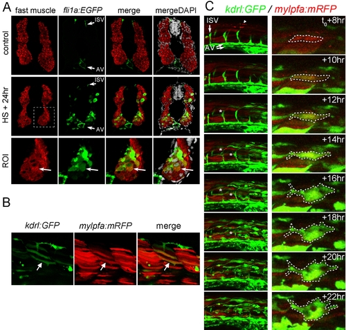
Fast skeletal muscle expresses ectopic endothelial genes following Etv2 overexpression. (A) Immunostained sections through the trunk of 48 hpf hsp70l:etv2/fli1a:EGFP embryos that were untreated (control) or heat shocked at 24 hpf (HS+24 h). Sections were stained for GFP and fast muscle myosin. Nuclei are stained with DAPI in the mergeDAPI panels. fli1a:EGFP is normally expressed in the intersomitic vessels (ISVs) and axial vessels (AVs) of control sections. However, following heat shock, many fast muscle myosin positive cells were also GFP positive (A). ROI is the region of interest highlighted by the dashed box in each panel. One section from 20 different embryos was observed for each treatment group with similar results within each group. (B) Confocal projection images of a kdrl:GFP+ and mylpfa:mRFP+ double positive muscle fiber (arrow) in a living embryo 12 h post–heat shock. (C) Time lapse imaging of the trunk (left column) and at the single cell level (right column) of a mylpfa:mRFP/hsp70l:etv2/kdrl:GFP triple transgenic embryo beginning at 8 h post–heat shock (t0+8 h). Heat shock occurred at 24 hpf. A few Etv2-mCherry+ nuclei are present in the first panel (arrowhead). The normal GFP+ intersomitic vessels (ISVs) and axial vessels (AVs) are labeled. mylpfa:mRFP labels fast muscle fibers in red. In the trunk, GFP expression first appears in muscle fibers between t0+8 h to t0+10 h and progresses in an caudal to rostral wave. mRFP+ fibers induce GFP expression and then soon switch off mRFP expression. ISV sprouts appear to apoptose and regress (asterisks). At the single cell level, mRFP+ fibers become GFP+ and then change morphology, a single cell is highlighted by a dashed outline in the right column. |