
Fig. 6
- ID
- ZDB-FIG-130826-72
- Publication
- Luo et al., 2013 - Compensatory Role of Inositol 5-Phosphatase INPP5B to OCRL in Primary Cilia Formation in Oculocerebrorenal Syndrome of Lowe
- Other Figures
- All Figure Page
- Back to All Figure Page
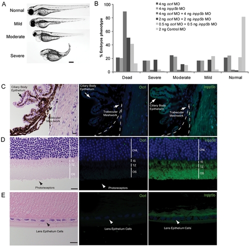
INPP5B and OCRL act synergistically in eye development. (A) Representative phenotypes of morphants co-injected with ocrl and inpp5b MO. Scale bar 250 micron. (B) Dose-dependent effect of two morpholinos in zebrafish. Control ocrl and inpp5b MO at indicated doses were injected into zebrafish embryos, and different grades of phenotypes were quantified at 48 hpf (N >250 embryos per group). (C) Transgenic mOcrl-/-:mInpp5b-/-:hINPP5B+/+ mouse eyes were sectioned and stained by H&E, anti-OCRL antibody (green) or anti-INPP5B antibody (green), and DAPI (blue). Staining of trabecular meshwork cells in vesicular pattern (arrow), dash line indicates border of cornea and iris. Scale bar 10 micron. (D) Photoreceptor cells (arrow) from the transgenic mouse eye section stained with H&E, anti-OCRL antibody (green) or anti-INPP5B antibody (green), and DAPI (blue). Scale bar 10 micron. (E) Lens epithelial cells (arrow) stained with H&E, anti-OCRL antibody (green) or anti-INPP5B antibody (green), and DAPI (blue). Scale bar 10 micron. |
| Fish: | |
|---|---|
| Knockdown Reagents: | |
| Observed In: | |
| Stage: | Long-pec |