
Fig. 5
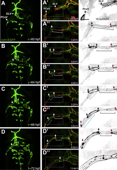
PCeV Forms through Cell Rearrangement and Cell-Splitting Events Still pictures of a time-lapse movie showing the formation of the PCeV (posterior (caudal) cerebral vein). (A)–(D) show a dorsal view (anterior up) of the head vasculature (green) at 60–72 hpf of a Tg(kdrl:EGFP)s843 zebrafish embryo. The fusing PCeV and DLV (dorsal longitudinal vein) are marked with white arrows. (A2)–(D3) show close-up of the PCeV fusion at time points corresponding to (A)–(D) of a transgenic embryo Tg(fliep:GFF)ubs3,(UAS:mRFP),(UAS:VE-cadherinΔC-EGFP)ubs12. Right panel shows a close-up of VE-cadherin-EGFP alone (black). Two lumenized sprouts approach each other led by single tip cells (A2, white arrows). A spot of VE-cadherin-EGFP is visible upon contact (A32, white arrow). The spot becomes a ring as the cell contact surface expands (B2 and B3, white/black arrows show ring edges). Transcellular lumen forms in both tip cells (B2–C3, white and blue bars). When the lumen is perfused, the new junctional ring approaches junctions of following cells and contacts them (C2 and D3, red arrows), indicating cell rearrangements that transform the vessel into a multicellular tube. Scale bars, 20 μm. See also Movie S5. |
Reprinted from Developmental Cell, 25(5), Lenard, A., Ellertsdottir, E., Herwig, L., Krudewig, A., Sauteur, L., Belting, H.G., and Affolter, M., In Vivo analysis reveals a highly stereotypic morphogenetic pathway of vascular anastomosis, 492-506, Copyright (2013) with permission from Elsevier. Full text @ Dev. Cell