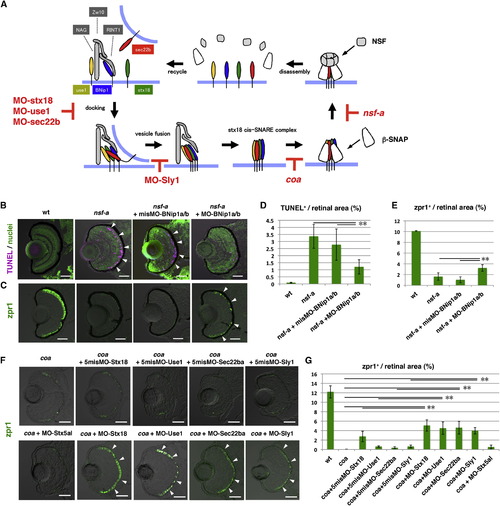
The Syntaxin-18 cis-SNARE Complex Is Required for BNip1-Dependent Apoptosis(A) Molecular mechanism underlying vesicular fusion to the ER. Interactions among syntaxin-18 (green), BNip1 (blue), Use1 (yellow), and Sec22b (red) initiate the fusion of transport vesicles to the ER membrane. Three accessory proteins, Zw10, NAG, and RINT1, play a role in the tethering of transport vesicles. After membrane fusion occurs, β-SNAP binds to the cis-SNARE complex and recruits NSF. NSF promotes disassembly of the cis-SNARE complex. In the absence of β-SNAP or NSF activity, the syntaxin-18 cis-SNARE complex accumulates. MOs against syntaxin-18, Use1, Sec22b, and Sly1 inhibit the formation of the syntaxin-18 cis-SNARE complex, even in the absence of β-SNAP or NSF activity. (B) TUNEL staining (magenta) of 72 hpf WT, nsf-a mutant, and nsf-a mutant retinas injected with misMO-BNip1a/b, and a mixture of MO-BNip1a and MO-BNip1b. Nuclei were counterstained with SYTOX Green (green). Arrowheads indicate apoptosis in the ONL. (C) Zpr1 labeling (green) of 84 hpf WT, nsf-a mutant, nsf-a mutant retinas injected with misMO-BNip1a/b, and a mixture of MO-BNip1a and MO-BNip1b. Arrowheads indicate rescued photoreceptors. (D and E) Percentage of TUNEL-positive (D) and zpr1-positive areas (E) relative to the total retinal area in the experiments shown in (B) and (C), respectively. Green and black bars indicate the means and SDs, respectively. **p < 0.01. (F) Zpr1 labeling (green) of coa mutant and coa mutants injected with MO-syntaxin-18, MO-Use1, MO-Sec22ba, MO-Sly1, their 5misMOs, and MO-syntaxin 5al. Arrowheads indicate rescued photoreceptors. (G) Percentage of zpr1-positive area relative to the total retinal area in the experiments shown in (F). Green and black bars indicate the means and SDs, respectively (**p < 0.01). The numbers of retinal sections used in the experiments shown in (D), (E), and (G) and p values for the t test are shown in Table S2. Scale bars, 50 μm. See also Figure S5.
|
