
Fig. 1
- ID
- ZDB-FIG-130813-5
- Publication
- Eames et al., 2013 - FishFace: interactive atlas of zebrafish craniofacial development at cellular resolution
- Other Figures
- All Figure Page
- Back to All Figure Page
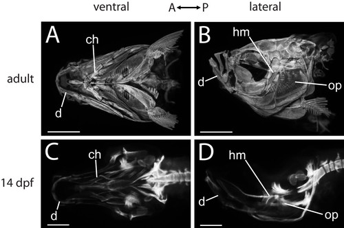
OPT data provide an overview of zebrafish craniofacial skeletal anatomy. Ventral (A, C) and lateral (B, D) views of zebrafish heads from OPT data demonstrate the decrease in complexity of skeletal elements when earlier specimens are compared with older specimens. While the adult head is covered by bones (A, B), only a small number of ossifications are visible in the ventral region of the 14 dpf zebrafish craniofacial skeleton (C, D). Representative skeletal elements are indicated. Abbreviations: A = anterior; ch = ceratohyal; d = dentary; dpf = days post-fertilization; hm = hyomandibula; op = opercle; P = posterior. Scale bars: A,B = 1 mm; C,D = 200 μm. |