
Fig. 4
- ID
- ZDB-FIG-130808-54
- Publication
- Lee et al., 2013 - An exclusively mesodermal origin of fin mesenchyme demonstrates that zebrafish trunk neural crest does not generate ectomesenchyme
- Other Figures
- All Figure Page
- Back to All Figure Page
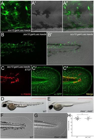
Neural crest cells do not contribute to fin mesenchyme. (A-B2) Lateral trunk (A-A2) and tail region (B,B2) of 48-hpf sox10:gal4; uas:kaede transgenic embryos. Kaede protein fluorescence (green) is observed in melanophores (A-A2), nascent dorsal root ganglia and spinal nerves (A), as well as in the fin (B,B2). (C-C2) Immunofluorescent labelling of Kaede (red, C), eGFP (green, C2) and merged image (C2) of the fin of a sox10:gal4; uas:kaede; ET37 transgenic embryo. (D-G) Overviews (D,E) and Nomarski images (F,G) of 48-hpf wild-type (WT) (D,F) and mos-/-; mob-/- (E,G) embryos showing loss of pigment but presence of a fully formed medial fin (E) with fin mesenchyme cells (G) in the double mutant. Red brackets indicate the region used for quantifying fin mesenchyme cells in H. (H) Quantification of fin mesenchyme cells in 12 WT and 12 mos-/-; mob-/- embryos at 48 hpf. No significant differences (n.s.) were observed (two-tailed Student’s t-test). Bars indicate the mean. |
| Gene: | |
|---|---|
| Antibodies: | |
| Fish: | |
| Anatomical Terms: | |
| Stage: | Long-pec |