
Fig. 2
- ID
- ZDB-FIG-130805-1
- Publication
- Shin et al., 2012 - Intrinsic and extrinsic modifiers of the regulative capacity of the developing liver
- Other Figures
- All Figure Page
- Back to All Figure Page
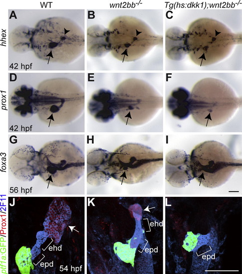
Loss of Wnt/β-catenin signaling does not affect the formation of the extrapancreatic duct. (A-F) hhex (A-C) and prox1 (D-F) expression in the liver-forming region (arrows) at 42 hpf was further reduced in Wnt/β-catenin signaling deficient embryos compared to wnt2bb mutants. However, hhex expression in the dorsal pancreas appeared unaffected (A-C, arrowheads). (G-I) foxa3 expression was examined to reveal the overall gut morphology. A small liver bud formed in wnt2bb mutants at 56 hpf (H), whereas it did not form in Wnt/β-catenin signaling deficient embryos (I). Arrows point to the liver-forming region. Dorsal views, anterior to the left. (J-L) The extrahepatic and extrapancreatic ducts (ehd and epd) were revealed by mAb 2F11 staining, the exocrine pancreas by Tg(ptf1a:GFP) expression, and the liver by Prox1 expression (arrows). Both the liver and the extrahepatic duct failed to form in Wnt/β-catenin signaling deficient embryos (L). Ventral views, anterior up. Scale bars, 100 µm. |
| Genes: | |
|---|---|
| Antibodies: | |
| Fish: | |
| Condition: | |
| Anatomical Terms: | |
| Stage: | Long-pec |
| Fish: | |
|---|---|
| Observed In: | |
| Stage: | Long-pec |
Reprinted from Mechanisms of Development, 128(11-12), Shin, D., Weidinger, G., Moon, R.T., and Stainier, D.Y., Intrinsic and extrinsic modifiers of the regulative capacity of the developing liver, 525-535, Copyright (2012) with permission from Elsevier. Full text @ Mech. Dev.