|
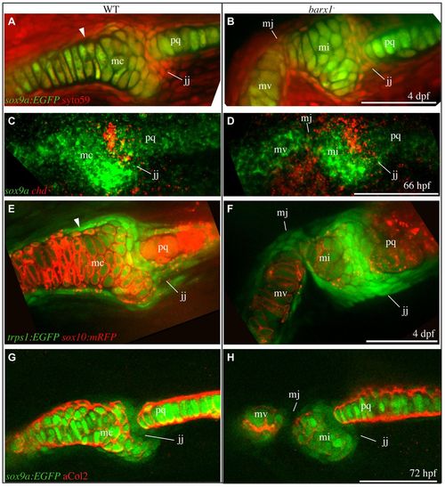
Ectopic joint cells are present in barx1 mutant zebrafish. (A,B) All cells were stained with SYTO59 (red), and chondrocytes transgenically labeled with EGFP (sox9a:EGFP, green) in 4 dpf animals. (C,D) 66 hpf larvae were subjected to in situ hybridization to label cartilage (sox9a, green) and joint cells (chd, red). (E,F) Live larvae with chondrocyte cell membranes transgenically labeled with RFP (sox10:mRFP, red) and joint region cells labeled with EGFP (trps1:EGFP, green) were imaged. (G,H) 72 hpf larvae with chondrocytes transgenically labeled with EGFP (sox9a:EGFP, green) were stained with anti-collagen type II antibody (red). Skeletal elements are oriented and labeled as in Fig. 1F. Arrowhead indicates boundary between distinct chondrocyte populations. All images are single confocal sections. Scale bars: 50 μm.
|