
Fig. 1
- ID
- ZDB-FIG-130726-27
- Publication
- Head et al., 2013 - Activation of canonical Wnt/beta-catenin signaling stimulates proliferation in neuromasts in the zebrafish posterior lateral line
- Other Figures
- All Figure Page
- Back to All Figure Page
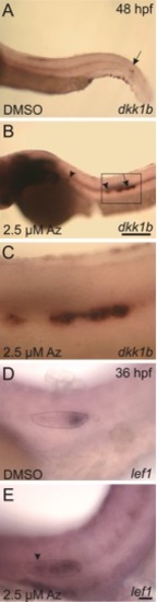
Expression of the Wnt targets dkk1 and lef1 is upregulated in the lateral line primordium in response to treatment with 2.5 μM Az beginning at 24 hpf. A–C: In situ hybridization with a probe against dkk1 shows normal expression in control, DMSO-treated embryos at 48 hpf (A) restricted to a narrow domain of the lateral line primordium (arrow), which has migrated almost to the tail. In contrast, in fish at 48 hpf treated with Az (B), the primordium has only migrated to approximately halfway along the yolk-extension, and dkk1 is expressed throughout the primordium (arrow) and remains elevated in deposited neuromasts (arrowheads). The boxed area is shown at higher magnification in C. D,E: In situ hybridization in DMSO-treated control fish at 36 hpf (D) shows normal expression of lef1 at the leading edge of the primordium (outlined with the dotted line). In Az-treated fish at 36 hpf, lef1 is expressed throughout the primordium and in the rosette about to be deposited (arrowhead). Scale bars: A,B = 250 μm; C–E = 25 μm. |