|
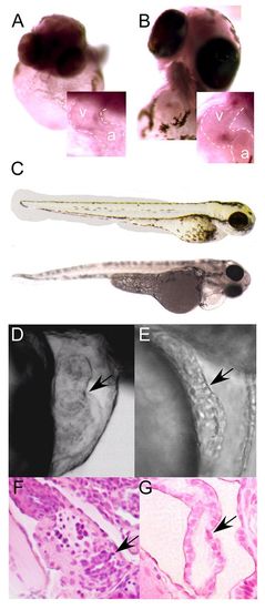
MicroRNA 21 expression and knockdown. (A,B) In situ hybridization with mature miR-21 locked nucleic acid (LNA) probe at 48 (A) and 72 (B) hpf demonstrates expression in zebrafish atrioventricular canal (AVC) at 48 hours (A, inset) and staining of atrioventricular valve (AV) and outflow tract (B, inset). (C) miR-21 knockdown does not perturb the normal body plan or axis at 72 hpf. Control, top; morphant, bottom. (D,E) Mismatch MO control-injected embryos show normal AV ring constriction (D, arrow) at 48 hpf, whereas miR-21 knockdown results in loss of the normal AV constriction (E, arrow). There is also failure of normal cardiac looping, as well as pericardial edema. (F,G) Hematoxylin and Eosin stained sections at day 5 post-fertilization demonstrate normal AV in controls (F, arrow) but failure of AV development in miR-21 knockdown embryos (G, arrow). In all images, the atrial chamber (a) is below and to the right and the ventricle (v) is above and to the left.
|